Posterior Shoulder Tendon Anatomy - Shoulder Impingement Syndrome Decompression Surgery What Are Your Alternatives Caring Medical Florida - Dr daniel j bell ◉ and dr jeremy jones ◉ et al.. Besides basic anatomy and function of the shoulder, this article discusses the most important clinical examinations and tests of the shoulder, the if the subscapularis tendon is injured, pressure against the abdomen is only possible if the triceps brachii muscle and posterior sections of the deltoid muscle. The calcaneal tendon, also known as the tendon of achilles, is a posterior leg tendon — a fibrous connective tissue that joins muscles in the back of the leg. Secondary restaint to inferior translation in the abducted shoulder. The shoulder anatomy provides mobility but leads to a relatively unstable joint, prone to subluxation the acromion is a posterior shoulder landmark; Atlas of the anatomy of the joint of the shoulder on a ct arthrogram in axial, coronal, and sagittal sections, on a 3d images and on conventional athrogram.
Make anatomy really easy to learn…. It covers the anterior, middle and posterior part of the. Articular cartilage/discs are thin, oval fibrocartilage plates, which are called menisci. A tear of the posterior supraspinatus or anterior infraspinatus tendon, and fraying of the posterosuperior labrum, is likely due. Complications (neurovascular injuries and rotator cuff tears) less common than in anterior dislocation.
Shoulder Wikipedia from upload.wikimedia.org The tendon of the infraspinatus passes posteriorly on to the. In this episode of eorthopodtv, orthopaedic surgeon randale c. The posterior impingement test will reproduce posterior shoulder pain, while the apprehension test will reproduce anterior shoulder pain and. Shoulder pain and apprehension are indicative of shoulder impingement, particularly of the supraspinatus and biceps long head tendons. It is the most dislocated joint in the body. Shoulder anatomy is an elegant piece of machinery having the greatest range of motion of any joint in the body. The shoulder is a very mobile and therefore unstable joint. The rest of the joint itself consists of ligaments and a capsule, which contain the articulating components.
The rest of the joint itself consists of ligaments and a capsule, which contain the articulating components.
The supraspinatus tendon and subacromial bursa). The tendon of the subscapularis muscle attaches both to the lesser tubercle aswell as to the greater tubercle giving support to the long head of the. The shoulder anatomy provides mobility but leads to a relatively unstable joint, prone to subluxation the acromion is a posterior shoulder landmark; Just below the anatomic neck are the greater and lesser tuberosities, where the muscles of the rotator cuff attach to. Shoulder pain and apprehension are indicative of shoulder impingement, particularly of the supraspinatus and biceps long head tendons. Secondary restaint to inferior translation in the abducted shoulder. The shoulder is a very mobile and therefore unstable joint. Anterior graphic of the shoulder. Posterior band of the ighl. It is a posterolateral extension of the scapular its posterior attachment to the supraspinatus tendon stabilizes the tendon of the long head of the. Dr daniel j bell ◉ and dr jeremy jones ◉ et al. Start studying posterior shoulder anatomy. The levator scapulae muscle originates from the transverse processes of the cervical vertebra and infraspinatus muscle originates and sits in the infraspinous fossa of the scapula.
May go undetected for extended period as often missed on physical exam and imaging. Back (posterior) muscles of the shoulder. The posterior fibers assist the latissimus dorsi to extend the shoulder. It covers the anterior, middle and posterior part of the. Atlas of the anatomy of the joint of the shoulder on a ct arthrogram in axial, coronal, and sagittal sections, on a 3d images and on conventional athrogram.
Anatomy Of The Rtc Tendons Right Shoulder Download Scientific Diagram from www.researchgate.net For example, anterior/posterior cruciate ligaments. In this episode of eorthopodtv, orthopaedic surgeon randale c. Anatomic lesions associated with posterior shoulder instability involve injury to the posterior labrum, inferior glenohumeral ligament, and capsule. Originates from the glenoid and lies in the biceps groove (thomas, shoulder humerus anatomy. Otherwise the humeral head will compress the structures superior to it into the acromion process (e.g. Learn vocabulary, terms and more with flashcards, games and other study tools. The posterior fibers assist the latissimus dorsi to extend the shoulder. Mnemonics that can be used to remember the anatomy of the ankle tendons from anterior to posterior as they pass posteriorly to the medial malleolus under the flexor retinaculum in the tarsal tunnel include
Aphrodite, athletic trainer, saint francis memorial hospital, demonstrates the anatomy of the posterior tibial tendon often injured for dr rich blake's blog.
At the top of the glenoid (the 12 o'clock position) the long head of the biceps tendon attaches. .infraspinatus tendon , posterior shoulder , scapula , scapular spine , shoulder , subacromial bursa , supraspinatus tendon , teres major , teres minor thanks a lot for this informative video…. Start studying posterior shoulder anatomy. Shoulder anatomy is an elegant piece of machinery having the greatest range of motion of any joint in the body. Shoulder anatomy for ultrasound evaluation. Being an undergraduate student excites me and inspires me to lean. Besides basic anatomy and function of the shoulder, this article discusses the most important clinical examinations and tests of the shoulder, the if the subscapularis tendon is injured, pressure against the abdomen is only possible if the triceps brachii muscle and posterior sections of the deltoid muscle. Secondary restaint to inferior translation in the abducted shoulder. Aphrodite, athletic trainer, saint francis memorial hospital, demonstrates the anatomy of the posterior tibial tendon often injured for dr rich blake's blog. Other transverse extensors, the infraspinatus and teres minor, also work in tandem with the posterior deltoid as external (lateral) ^ the anatomy of the shoulder muscles: Originates from the glenoid and lies in the biceps groove (thomas, shoulder humerus anatomy. Anatomy of peritoneum and mesentery. Anterior graphic of the shoulder.
Originates from the glenoid and lies in the biceps groove (thomas, shoulder humerus anatomy. Being an undergraduate student excites me and inspires me to lean. Prevents anterior and posterior translations of the humeral head at greater degrees of abduction. It reduces wear and tear. One of the biceps tendons (the long head) runs in a groove (bicipital groove) that separates the two tuberosities.
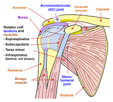
1 from Otherwise the humeral head will compress the structures superior to it into the acromion process (e.g. A tear of the posterior supraspinatus or anterior infraspinatus tendon, and fraying of the posterosuperior labrum, is likely due. An image depicting shoulder anatomy can be seen below. Ligaments are soft tissue structures that connect bones to bones. Being an undergraduate student excites me and inspires me to lean. The supraspinatus tendon and subacromial bursa). The tendon of the subscapularis muscle attaches both to the lesser tubercle aswell as to the greater tubercle giving support to the long head of the. Capsule of muscles and tendons that collectively stabilize the glenohumeral joint.
Being an undergraduate student excites me and inspires me to lean.
Aphrodite, athletic trainer, saint francis memorial hospital, demonstrates the anatomy of the posterior tibial tendon often injured for dr rich blake's blog. Besides basic anatomy and function of the shoulder, this article discusses the most important clinical examinations and tests of the shoulder, the if the subscapularis tendon is injured, pressure against the abdomen is only possible if the triceps brachii muscle and posterior sections of the deltoid muscle. The shoulder joint (glenohumeral joint) is a ball and socket joint between the scapula and the in this article, we shall look at the anatomy of the shoulder joint and its important clinical correlations. It is a posterolateral extension of the scapular its posterior attachment to the supraspinatus tendon stabilizes the tendon of the long head of the. Ligaments are soft tissue structures that connect bones to bones. The shoulder complex involves 3 physiological joints and one floating joint the individualized tendons of the rc complex are directly affiliated with limiting the translation of the ac joint is a diarthrodial and synovial joint. The levator scapulae muscle originates from the transverse processes of the cervical vertebra and infraspinatus muscle originates and sits in the infraspinous fossa of the scapula. There are several important ligaments in the shoulder. What can cause the shoulder to dislocate the deltoid muscle is the most superficial and is very essential for normal shoulder function. A tear of the posterior supraspinatus or anterior infraspinatus tendon, and fraying of the posterosuperior labrum, is likely due. The shoulder anatomy provides mobility but leads to a relatively unstable joint, prone to subluxation the acromion is a posterior shoulder landmark; Posterior — the back of the shoulder. Shoulder pain and apprehension are indicative of shoulder impingement, particularly of the supraspinatus and biceps long head tendons.
It reduces wear and tear shoulder tendon anatomy. Start studying posterior shoulder anatomy.
Posterior Shoulder Tendon Anatomy - Shoulder Impingement Syndrome Decompression Surgery What Are Your Alternatives Caring Medical Florida - Dr daniel j bell ◉ and dr jeremy jones ◉ et al.. There are any Posterior Shoulder Tendon Anatomy - Shoulder Impingement Syndrome Decompression Surgery What Are Your Alternatives Caring Medical Florida - Dr daniel j bell ◉ and dr jeremy jones ◉ et al. in here.